Joint pain and tendon dysfunction are among the most common barriers to sustained physical activity. Maintaining connective tissue health requires proactive nutritional, hormonal, and mechanical support, and injury recovery must be managed carefully to avoid impeding the repair process.
Joint & Tendon Support: Evidence-Based Protocols for Connective Tissue Health and Repair
Joint pain and tendon dysfunction are among the most common barriers to sustained physical activity. Maintaining connective tissue health requires proactive nutritional, hormonal, and mechanical support, and injury recovery must be managed carefully to avoid impeding the repair process.
Quick summary
- This page explains how we care for your joints and tendons.
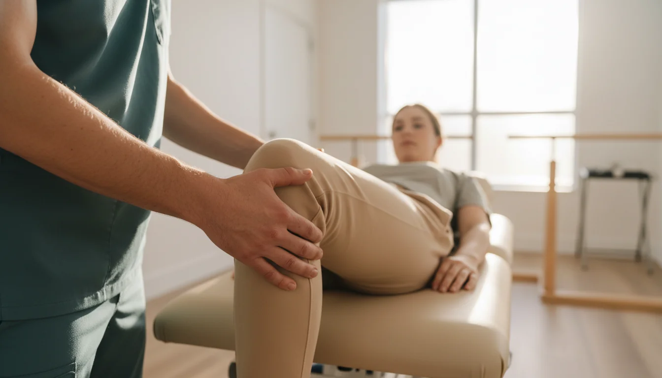
- We use exams and imaging to find the source of pain.
- Care may include nutrition, hormone checks, and physical therapy.
- Every plan is led by a licensed doctor and reviewed over time.
This content is for educational purposes only. It does not replace an in-person or telehealth consultation with a licensed clinician, and does not constitute medical advice or a treatment plan. All treatment decisions — including the use of hormones, peptides, supplements, or other agents — require individual clinical evaluation, laboratory confirmation, and licensed physician oversight. Do not self-administer any medication or compound based on information in this article.
Joint pain and tendon dysfunction are among the most common barriers to sustained physical activity. Unlike skeletal muscle — with abundant vascularity and regenerative capacity — tendons and articular cartilage are relatively avascular structures with limited self-repair potential. This biological reality means that maintaining connective tissue health requires proactive nutritional, hormonal, and mechanical support, and that injury recovery must be managed carefully to avoid impeding the repair process.
At Advanced Vitality Group, joint and tendon support programs are built on the available clinical evidence. This includes interventions with strong RCT support (collagen nutrition, progressive loading), those with moderate evidence (omega-3 fatty acids, vitamin D), and those with preclinical data and limited human trials (BPC-157) — with transparent communication about which is which.
The Biology of Tendons and Cartilage
Tendons
Tendons are composed primarily of Type I collagen organized in a hierarchical structure. Tenocytes — the cells responsible for tendon maintenance — have limited proliferative capacity and a poor direct blood supply, which is why tendon injuries heal slowly. Collagen synthesis by tenocytes requires mechanical loading as the primary stimulus, adequate protein substrate, vitamin C (for hydroxylation enzymes), and appropriate hormonal signaling including IGF-1.
Articular Cartilage
Articular cartilage is avascular, aneural, and has extremely limited regenerative capacity. Chondrocytes cannot replicate significant tissue loss once degradation has occurred. This makes prevention — through anti-inflammatory management, collagen nutrition, mechanical optimization, and correction of contributing biological deficiencies — the clinically viable strategy.
Collagen Nutrition: The Strongest Available Evidence
Collagen comprises approximately 70–80% of dry tendon weight and is the primary structural component of articular cartilage. The most clinically significant human trial in this area was a randomized crossover study by Shaw G et al. (American Journal of Clinical Nutrition, 2017), which demonstrated that 15 g of vitamin C-enriched gelatin consumed one hour before exercise significantly increased collagen synthesis (serum P1NP) compared to placebo. This established a practical, evidence-based protocol for supporting connective tissue repair through nutrition.
| Intervention | Mechanism | Evidence | Protocol |
|---|---|---|---|
| Hydrolyzed collagen peptides (15 g) + Vitamin C (500 mg) | Provides substrate (proline, glycine, hydroxyproline) and cofactor (vitamin C for hydroxylation) for collagen synthesis | RCT: increased P1NP vs. placebo (Shaw et al., 2017); meta-analysis: reduced joint pain (García-Coronado et al., 2019) | 1 hour before exercise/rehabilitation |
| Omega-3 fatty acids (EPA + DHA, 2–4 g/day) | Reduces prostaglandin E2 and pro-inflammatory cytokines in joint synovium | Meta-analyses of RCTs in RA and OA: reduced joint pain and morning stiffness | 2–4 g/day combined EPA+DHA |
| Vitamin D (correct documented deficiency) | VDRs in chondrocytes and synoviocytes; deficiency associated with cartilage loss in knee OA | Prospective cohort data (McAlindon et al., AnnIM, 1996); RCTs for deficiency correction | Correct confirmed deficiency to resolve insufficiency |
These are evidence-based nutritional interventions, not universal performance targets. Vitamin D targets, for example, should be determined in clinical context — not applied as fixed optimization values.
Progressive Loading: The Primary Treatment for Tendinopathy
The most consistent evidence-supported intervention for tendinopathy is progressive mechanical loading — not rest. The Alfredson eccentric loading protocol for Achilles tendinopathy (3×15 eccentric heel drops twice daily, 7 days/week) is supported by multiple RCTs demonstrating superiority over rest (Alfredson H et al., American Journal of Sports Medicine, 1998). Heavy isometric loading — 45-second holds at approximately 70% MVC — has demonstrated immediate analgesic effects in patellar tendinopathy and is appropriate in early rehabilitation phases when full eccentric loading is not yet tolerated (Rio E et al., BJSM, 2015).
Complete rest is not the evidence-based recommendation for most tendinopathies. Tendons require mechanical load to activate tenocytes, stimulate collagen synthesis, and drive proper remodeling. The key is appropriate loading dose — enough to stimulate adaptation without exceeding the tissue's current capacity.
BPC-157 for Joint and Tendon Health: Evidence and Limitations
BPC-157 has a substantial preclinical literature for musculoskeletal applications: animal studies demonstrate accelerated Achilles tendon repair, improved collagen organization, cartilage and bone protection, and VEGF-mediated angiogenesis in relatively avascular tissues. The proposed mechanisms — VEGF upregulation, GH receptor activation, fibroblast stimulation — are biologically plausible for connective tissue repair.
However, published human RCT data for joint and tendon indications remains limited. BPC-157 is not FDA-approved for any indication. At Advanced Vitality Group, BPC-157 protocols for joint and tendon applications are offered as investigational options with transparent disclosure of: the preclinical evidence base; the absence of published large-scale human RCTs; the regulatory status; and the compounding considerations involved. Patients receive this information before making any decision.
Inflammation and Joint Degradation
Chronic systemic inflammation — elevated hs-CRP, IL-6, and TNF-α — accelerates cartilage degradation, promotes synovial inflammation, and impairs tendon repair. Addressing systemic inflammaging (described in detail in our Inflammation Control program) creates a more favorable environment for joint preservation. Omega-3 fatty acids, Mediterranean dietary pattern adherence, and body weight optimization are among the most evidence-supported interventions for reducing synovial inflammation in non-inflammatory joint conditions.
Frequently Asked Questions
Scientific References
- Shaw G, et al. “Vitamin C-enriched gelatin supplementation before intermittent activity augments collagen synthesis.” American Journal of Clinical Nutrition. 2017;105(1):136–143.
- Alfredson H, et al. “Heavy-load eccentric calf muscle training for chronic Achilles tendinosis.” American Journal of Sports Medicine. 1998;26(3):360–366.
- Rio E, et al. “Isometric exercise induces analgesia and reduces inhibition in patellar tendinopathy.” BJSM. 2015;49(19):1277–1283.
- García-Coronado JM, et al. “Effect of collagen supplementation on osteoarthritis symptoms.” International Orthopaedics. 2019;43(3):531–538.
- Pevec D, et al. “Impact of BPC 157 on muscle healing.” Medical Science Monitor. 2010;16(3):BR81–88.
- McAlindon TE, et al. “Do antioxidant micronutrients protect against knee osteoarthritis?” Annals of Internal Medicine. 1996;125(5):353–359.
- Calder PC. “Omega-3 fatty acids and inflammatory processes.” Biochemical Society Transactions. 2017;45(5):1105–1115.
More in Performance & Recovery
View allPerformance Enhancement
Evidence-based evaluation of factors limiting exercise capacity and physical function.
Muscle Growth
Hormonal, nutritional, and training science for hypertrophy and sarcopenia prevention.
Energy Optimization
Metabolic, hormonal, thyroid, and mitochondrial causes of persistent low energy.
Post-Workout Recovery
Sleep, nutrition, hormonal balance, and inflammation for recovery speed.
Injury Repair
Growth factor support, collagen nutrition, and peptide therapy for tissue healing.
Joint & Tendon Support
You are hereCollagen synthesis, progressive loading, and connective tissue nutrition.
Schedule a consultation to evaluate your connective tissue health and build an evidence-based joint and tendon support protocol.